BIODEGRADATION OF POLYCYCLIC AROMATIC
HYDROCARBONS WITH ENTEROBACTER ASBURIAE
INTRODUCTION
Polycyclic aromatic hydrocarbons (PAHs) are of the
ubiquitous environmental pollutants found in different environmental
matrices at different concentrations (Nessim et al.
2011, Zou et al. 2011). Significant
accumulation of PAHs in the aquatic ecosystem had been caused by
anthropogenic inputs like oil spills, sea navigation, urban runoff,
water, and industrial wastes (Wu et al. 2011,
Westley et al. 2010). High concentrations of
PAHs are found in marine coastal environment near cities and
industrial plants (Opuene et al. 2007). PAHs
are a class of organic pollutants characterized by the fusion of two
or more aromatic rings composed of carbon and hydrogen atoms. These
compounds typically exist in the form of colorless, white, or
pale-yellow solid substances (Abdel-Shafy & Mansour 2016, Suman
et al. 2016). The spatial arrangement of the
aromatic rings can vary, including linear, angular, or clustered
configurations (Abdel-Shafy & Mansour 2016).
PAHs can be categorized based on the number of aromatic
rings they contain, resulting in two main groups: light-molecular
weight PAHs (LMW PAHs) with two or three rings and high-molecular
weight PAHs (HMW PAHs) with four or more rings. Depending on their
molecular weight, LMW PAHs are released into the atmosphere in gaseous
form, while HMW PAHs are found in particulate matter (Lee & Vu
2010). Furthermore, PAHs can also be classified based on their ring
structures. Alternant PAHs consist solely of fused six-carbon benzene
rings, whereas non-alternant PAHs, such as fluorene, include
six-carbon benzene rings fused with an additional ring containing
fewer than six carbon atoms (Gupte et al.
2016).
The presence of dense p electrons in the aromatic rings
contributes to the chemical persistence of PAHs, making them resistant
to nucleophilic attacks (Haritash & Kaushik 2009). In 1983, the
United States Environmental Protection Agency (USEPA) identified 16
PAHs as priority pollutants due to their high concentrations,
extensive exposure, recalcitrance, and toxicity (Zheng et al. 2018, Mojiri et al.
2019). PAHs exhibit low water solubility, low vapor pressure, and
high melting and boiling points, which vary depending on their
specific chemical structures (Lee & Vu 2010). As the molecular
weight of PAHs increases, their water solubility decreases, and they
become more lipophilic, rendering them more persistent compounds
(Okere & Semple 2012).
Anthracene is known to cause direct skin irritation and
skin sensitization in both animals and humans (Rengarajan et al. 2015). Chronic health effects associated
with PAH exposure include conditions like cataracts, kidney and liver
damage, respiratory issues, reduced immune function, lung dysfunction,
and symptoms resembling asthma (Abdel-Shafy & Mansour 2016).
Naphthalene can lead to the breakdown of red blood cells if inhaled or
ingested in high concentrations (Rengarajan et al.
2015). Fluorene is linked to adverse effects on both wildlife and
human health, exhibiting acute toxicity as well as potential mutagenic
and carcinogenic properties (Yu 2002).
Concern about PAHs has predominantly focused on their
carcinogenic properties (Boffetta et al. 1997,
Rubin 2001). Recently, there has been increased focus on the potential
effects of polycyclic aromatic hydrocarbons on human health.
Specifically, their ability to disrupt hormonal functions related to
reproduction and depress immune system function has come under
examination (Uppstad et al. 2011). These
concerns have prompted both the World Health Organization (WHO) (WHO
2006) and the United States Environmental Protection Agency (USEPA)
(U.S. EPA 2003) to formulate regulations for the protection of
drinking and source water systems in order to safeguard the populace
from such harmful pollutants, many of which are considered as
potential carcinogens. Degradation of PAHs in the environment
includes: biodegradation, photooxidation,
and chemical
oxidation adsorption to soil particles, leaching,
bioaccumulation (Tolosa et al. 1996). Each of
these processes affects individual PAHs in a different manner. This is
mainly due to the fact that each PAH has a unique structure and a set
of physical, chemical, and biological properties. On the other hand;
PAHs biodegradation studies focused on aerobic degradation.
Nevertheless, anaerobic degradation has been demonstrated (Haritash
& Kaushik 2009,Peng
etal. 2008). In
order for bacteria to degrade any given PAH, it must be made available
for uptake by the bacteria (Cerniglia2003,Dandie
et al. 2004,Fredslund
et al. 2008). PAHs become bioavailable
when they are in either the dissolved or the vapor phase.
Bioremediation is a technology that transforms relatively toxic
compounds to less hazardous forms using biological processes.
Polycyclic aromatic hydrocarbons degradation depends on several
factors including: the environmental conditions, number and type of
the microorganisms, nature and chemical structure of the chemical
compound being degraded. The PAHs- degrading microorganism includes
algae, bacteria, and fungi. It involves the breakdown of organic
compounds through biotransformation into less complex metabolites, and
through mineralization into inorganic minerals, H2O, CO2 (aerobic) or CH4 (anaerobic) (Abdel-Shafy & Mansour 2016).
This study aims to address the knowledge gap in
environmental pollution monitoring by investigating polycyclic
aromatic hydrocarbons (PAHs) in specific contaminated sites in Egypt
that have not been previously monitored for PAH pollution. The
hypotheses involve the potential existence of bacteria in the
contaminated water capable of effectively reducing polycyclic aromatic
compounds. The experimental procedure involves the collection of water
samples, isolation of bacteria, screening for the most effective
isolates, in vitro evaluation of their efficacy in reducing PAHs,
identification of the most effective isolates, and subsequent use of
the isolates for in vitro removal of PAHs. The anticipated outcomes
include the quantification of PAH concentrations in selected sites,
isolation of diverse bacterial isolates with PAH biodegradation
capabilities, characterization and identification of these isolates,
and conducting biodegradation assays to assess the selected bacterial
isolate's proficiency in degrading PAHs. The findings will contribute
to the understanding of microbial remediation strategies for
PAH-contaminated environments.
MATERIALS AND METHODS
Sampling and sampling sites:
industrial wastewater samples were collected in 2021 from three sites
in Kafr El-Sheikh Governorate, Egypt: Neamaa restaurant, Kitchener
Drain, and a Total gas station. The samples were collected manually
on-site and transported to the laboratory in iceboxes. Table I showed
sampling sites locations and coordinates.
Chemicals: the tested PAHs
active ingredients were anthracene (100.0 % purity), fluorene (99.1 %
purity) and naphthalene (99.8 % purity). The active ingredients were
supplied by AccuStandard Inc., USA. They were obtained from the
Central Laboratory at the Kafr El-Sheikh Company for Water and
Wastewater in Kafr El-Sheikh City, Egypt. Gradient grade acetonitrile
from Merck Millipore, Germany was used as the exchange solvent for
liquid chromatography. Methylene chloride (99.0 % purity) from
Sigma-Aldrich, USA was used to extract the PAHs.
Sample Preparation: two-liter
water samples were collected from each site in cleaned, sterilized,
solvent-washed glass bottles. The samples were transported to the
laboratory in ice containers. One liter of each sample was filtered
through fiberglass -Grade MGC Micro-Glass Fiber Filters, Munktell
Filtrak™ , CAT N°. 3.1103.024- to remove turbidity and debris. These
filtered samples were stored at 4°C prior to extraction and then
tested for PAH concentrations. The remaining one-liter samples were
incubated at 30°C for two weeks. After the incubation period, these
samples were tested using the same methods in order to detect
percentages of PAH biodegradation (Belal et al.
2018).
Media: three types of
enrichment media were used in this study as described by Brunner et al. (1980): minimal medium as mineral salt
liquid (MSL), minimal medium as mineral salt agar, and nutrient
agar.
Analysis of HPLC: approximately
1 L of each sample was serially extracted three times with 60.0 mL of
methylene chloride using a separatory funnel. The methylene chloride
extract was dried and concentrated to a volume of 1 mL. Then, 3.0 mL
of acetonitrile (the exchange solvent) was added and the extract
concentrated again to a final volume of 0.5 mL. The extract analytes
were separated by HPLC. Ultraviolet adsorption (UV) and fluorescence
detectors coupled to the HPLC system were used to quantitatively
measure the PAHs. (Hodgeson 1990).
High performance liquid chromatography (HPLC)
Quantitative determination of polycyclic aromatic
hydrocarbons (PAHs) was performed using a Dionex Ultimate 3000 high
performance liquid chromatography (HPLC) system (Dionex USA) at the
Central Laboratory, Kafr El-Sheikh Company for Water and Wastewater
in Kafr El-Sheikh, Egypt.
Isolation by enrichment culture
Enrichment cultures capable of degrading PAHs were
established using water samples collected from the three industrial
wastewater sites in Kafr El-Sheikh Governorate, Egypt. The samples
were tested to detect PAH concentrations. A 10 mL aliquot of each
sample was suspended in 90 mL of sterilized mineral salt medium
(MSL) in 500-mL bottles containing 1 μg/L of the PAH active
ingredients as the sole carbon source. These enrichment cultures
were incubated at 30°C and 150 rpm for 14 days. Next, a 10 mL
aliquot from each culture was transferred to fresh 90 mL MSL medium
with the same 1 μg/L PAH concentration. This subculture procedure
was repeated four times. After the final transfer, dilution series
of the enrichment cultures were prepared in glass tubes containing 9
mL MSL medium, to dilutions of 10-6. Then, 100
μL aliquots of each dilution were spread onto MSL medium plates
containing 1 μg/L PAHs using Drigalski spatulas. The inoculated
plates were sealed in polyethylene bags and incubated at 30°C for 7
days, monitoring for bacterial colony growth. Single colonies
appearing on the diluted plates were isolated by picking with
sterile inoculation needles. The isolates were further purified
using standard spatial streaking onto complex agar media. (Derbalah
& Belal 2008, Derbalah et al. 2008,
Massoud et al. 2008).
Screening of bacterial isolates using the viable count
technique
The isolated colonies underwent testing for their
ability to grow in MSL medium containing the PAHs standard at a
concentration of 1 µg/L. Two treatments were employed: one with the
medium and PAHs standard, and the other with only the medium as a
control (without PAHs). The cultures were agitated at 150 rpm and
maintained at a temperature of 30 °C for a duration of 14 days.
Screening of bacterial isolates with superior PAH-degrading
capabilities was conducted based on the viable count of these
isolates. Serial dilution tubes up to 1 * 10-6
were prepared from each culture, and a 100 µL volume from each tube
was spread onto plates of plate count agar medium using a sterilized
Drigalski triangle. The plates were then incubated at 30° C for 2
days, and the viable count of each strain was determined using a
colony counter. For further identification, single colonies of the
selected bacterial isolate (determined to be most proficient in
degrading PAHs) were isolated by picking the colonies with a sterile
needle. Subsequently, these colonies were inoculated into fresh
plates and slants for identification purposes, following the
procedures outlined in Derbalah & Belal (2008), Derbalah et al. (2008), and Massoud et
al. (2008).
Identification
The bacterial isolates capable of degrading selected
PAHs were identified through the examination of morphological and
biochemical characteristics, following the criteria established by
Gordon et al. (1973), Krieg & Holt
(1984), Chun et al. (2001), Logan (2005), and
Sharma et al. (2015). Additionally,
identification was confirmed using the 16S rRNA method outlined by
Boye et al. (1999).
Morphological and cultural characterization of the
selected:
. Bacterial
isolates were examined for their cell shape, motility studies, and
gram reaction.
. Purified
cultures at log phase after 72 h were microscopically examined for
the cell morphological characters.
. The 72-h grown
isolates were microscopically examined using cavity slide for
bacterial motility.
. Gram
staining was conducted following the procedure outlined by
Rangaswami & Bagyaraj (1993).
Biochemical characterization of the selective isolates:
. The
isolate was streaked onto trypticase soy agar medium and then
incubated at 37° C for 48 hours, following the method described by
Cappuccino & Sherman (1996). After the incubation period, 2-3
drops of a 1 % P-aminodimethylaniline oxalate solution were applied
to the streaked area. The plates were observed for a color change,
transitioning from pink to maroon, and ultimately to purple within
30 seconds, signifying a positive reaction.
. A
loopful of a 24-hour-old culture of the isolate was transferred to a
glass test tube containing 0.5 mL of distilled water. It was
thoroughly mixed with 0.5 mL of a 3 % hydrogen peroxide solution,
and the mixture was observed for effervescence, following the method
outlined by Gerhardt et al. (1981).
. Urease test
was performed on 5 mL of urea broth (20 g/L) in test tubes
containing phenol red (0.012 g/L), pH 6.8 as the pH indicator
(Cappuccino & Sherman 1996). The cultures were transferred into
the sterilized urea broth and incubated for 24 h. The development of
red color indicates a positive reaction.
. The selective isolate was inoculated into test tubes
having Simmons citrate agar medium and incubated for 48 h at 35°C
(Seeley & Vandemark 1981). Simmons citrate agar contains citrate
as only carbon and energy source. The presence of growth and change
of color from green to blue due to pH change indicated positive
reaction.
. The
isolate was inoculated into sterilized glucose tryptone broth in
test tubes (Seeley & Vandemark 1981). After 48 h of incubation,
0.3 mL of Kovac’s reagent was added and mixed well. The reddening of
the alcohol layer within few minutes indicates indole
production.
. A fresh pure
culture of the test culture was grown for 48 hours at 35°C in MR-VP
medium. After incubation, 5 drops of a 0.05 % methyl red solution
(per 2.5 ml) were added to the culture. The MR-positive shows a red
coloration as a result of high acid production and a decrease in the
pH of the culture medium to 4.4. The MR-negative culture has a
yellow color indicating a less acidic medium. (McDevitt 2009).
a fresh
pure culture of the test culture was grown in MR-VP-broth for 48
hours at 37°C and Barritt’s reagents A and B were added. VP-positive
shows red coloration on top of the culture as a result of Diacetyl
reacts with peptone in the medium, to form a pinkish red-colored
product, whereas VP-negative has a yellowish color. (McDevitt
2009).
Molecular characterization of the selective isolate by
polymerase chain reaction:
The most efficient bacterial isolates regarding PAHs
degradation were identified using 16S rRNA as described by Boye et al. (1999). DNA was extracted using protocol
of GeneJet genomic DNA, purified, and polymerase chain reaction
(PCR) was made by using Maxima Hot Start PCR Master Mix
(Thermo).
Biodegradation assay of polycyclic aromatic
hydrocarbons
An experiment was conducted to assess the
biodegradation capabilities of three polycyclic aromatic
hydrocarbons (PAHs) – Anthracene, Fluorene, and Naphthalene – using
the most proficient bacterial isolate capable of degrading these
compounds. An initial concentration of 5 ppb for each of the three
PAHs was introduced into MSL medium and inoculated with a bacterial
cell suspension from the most proficient isolate, with an inoculum
size of 106 cfu/ml. A control experiment was
established without the inoculation of a biological agent to monitor
abiotic losses. A duplicate experiment was performed to evaluate
biological degradation. All experiments were incubated at pH 7 and
30 °C for 14 days. Average concentrations, standard deviations, and
removal averages were calculated for each experiment. The
methodology employed aligns with that described by Belal et al. (2018).
Toxicity assessment of biodegradation byproducts
The toxicity bioassay for byproducts resulting from
the biodegradation of PAH compounds was conducted on aqueous
solutions after a 14-day incubation period with Enterobacter asburiae (Isolate N1). A bioassay test was performed using Bacillus subtilis, a gram-positive bacterium
chosen for its sensitivity to PAHs, serving as a suitable indicator
to assess the remaining toxicity of PAHs following treatment with
the tested microbial isolate. The selection of Bacillus subtilis as the target organism was
based on its demonstrated sensitivity to PAHs, as indicated by
Wulandari et al. (2021). To determine
toxicity, the inhibition zone in the growth of B.
subtilis was recorded and compared to the control treatment
(untreated). Bacillus subtilis was treated
with supernatants that had been incubated with the tested bacterial
isolate for 14 days. The test was standardized as follows: Nutrient
agar medium was poured into Petri dishes (9 cm in diameter, 15
ml/dish). After solidification, 100 μl of B.
subtilis (108 c.f.u/ml) was evenly spread
onto the nutrient agar plate using sterilized glass beads. Wells (5
mm in diameter) were punched in each plate. Subsequently, 50 μl of
the supernatant was added to the punched holes (5 mm in diameter) in
the nutrient agar medium. For the three PAHs compounds, each
incubated with Enterobacter asburiae (Isolate
N° 1), broth cultures were filtered through a sterile membrane
filter (0.2 μm). Conversely, a volume of 50 μl of sterilized liquid
medium was placed in the punched holes and used as a control
treatment. The experiments were conducted in triplicate. The plates
were incubated at 30 °C until full growth of the control treatments
was achieved (2 days). The diameter (mm) of the inhibition zone
surrounding each hole, where the growth of Bacillus subtilis was inhibited, was recorded.
Toxicity was determined as a percentage of inhibition in the growth
of the tested bacteria compared to the control treatment. Similar
bioassay protocols for assessing metabolite toxicity were employed
by Massoud et al. (2008), Hauka et al. (2014), Derbala & Belal (2008), and
Derbalah et al. (2008).
RESULTS AND DISCUSSION
This study conducted an analysis of three PAH
(Polycyclic Aromatic Hydrocarbon) compounds present in water samples
collected from three distinct sites in Kafr El-Sheikh governorate,
Egypt. The selected sites were Neamaa restaurant, Kitchener Drain, and
Total gas station. The compounds under investigation comprised
Naphthalene, Anthracene, and Fluorine. Table II presents the data,
revealing variable concentrations of PAHs in the water samples.
Table II shows the concentration (in ppb) of three
PAHs compounds - Anthracene, Fluorene, and Naphthalene - in water
samples collected from three industrial wastewater sources - Kitchener
drain, Neamaa restaurant, and total gas station - before and after
incubation period. It also shows the degradation percentage of these
compounds. Before incubation, the concentrations ranged from 0.424 to
253.108 parts per billion (ppb). The highest recorded value was
253.108 ppb for Anthracene found at the Total gas station site. In
contrast, the lowest recorded concentration was 0.424 ppb for Fluorene
detected in the Kitchener Drain samples. For Anthracene, there is a
high degradation percentage in all three sources after the incubation
period, ranging from 99.54 % in Kitchener drain to 100 % in Neamaa
restaurant and total gas station samples. This indicates that
Anthracene was efficiently degraded in the wastewater from all
sources. Fluorene shows varying degradation percentages - 46.23 % in
Kitchener drain, 100 % in Neamaa restaurant, and 100 % in total gas
station. While it was completely degraded in samples from Neamaa and
the gas station, the Kitchener drain sample retained over 50 % of
initial Fluorene after incubation. Naphthalene degradation ranges from
60.12 % in the gas station sample to 90.68% in the Kitchener drain.
The Neamaa restaurant sample shows 80.9 % reduction. While degradation
is occurring, Naphthalene persists after incubation most out of the
three PAHs across the samples. Additional measures may be required to
improve removal of this recalcitrant compound. Overall, the results
demonstrate the efficiency of degradation of PAH contaminants in
wastewater during the incubation period. However, persistence of
compounds like Fluorene and Naphthalene in some samples highlight the
need to further optimize treatment conditions.
Figures 1 and 2 shows chromatograms of three PAHs
compounds - Anthracene, Fluorene, and Naphthalene - in water samples
collected from Kitchener drain before and after incubation period
respectively, figures 3 and 4 shows chromatograms of three PAHs
compounds - Anthracene, Fluorene, and Naphthalene - in water samples
collected from Neamaa restaurant before and after incubation period
respectively and figures 5 and 6 shows chromatograms of three PAHs
compounds - Anthracene, Fluorene, and Naphthalene - in water samples
collected from total gas station before and after incubation period
respectively.
Fig.1.- Chromatogram of three PAHs compounds - Anthracene,
Fluorene, and Naphthalene - in water sample collected from Kitchener
drain before incubation period
Fig. 2. - Chromatogram of three PAHs compounds - Anthracene,
Fluorene, and Naphthalene - in water sample collected from Kitchener
drain after incubation period.
Fig. 3. - Chromatogram of three PAHs compounds - Anthracene,
Fluorene, and Naphthalene - in water sample collected from Neamaa
restaurant before incubation period
Fig. 4.- Chromatogram of three PAHs compounds - Anthracene,
Fluorene, and Naphthalene - in water sample collected from Neamaa
restaurant after incubation period
The varying degradation percentages of PAHs compounds
across the three industrial wastewater sources indicate that the
efficacy of bacterial remediation is impacted by site-specific
conditions. As noted by Gallego et al. (2001),
the composition and complexity of the microbial population, presence
of additional contaminants, and physico-chemical properties of water
can all affect bioremediation. Kitchener drain water likely contained
high organic content which can inhibit microbial activity and lowered
Anthracene and Naphthalene degradation despite adequate bacterial
populations (Yu et al. 2005). The Neamaa
restaurant wastewater, on the other hand, showed near complete PAHs
degradation, suggesting favorable microbial community and optimum
growth conditions (pH, temperature, nutrients etc.) (Chang et al. 2002). The differences also highlight that
while bacteria may efficiently metabolize some PAHs like Anthracene,
recalcitrant compounds like Naphthalene persist due to their complex
chemical structure. According to Samanta et al.
(2002), the presence of alkyl side chains inhibits enzyme
activities during microbial metabolism. Pre-exposure to low
Naphthalene levels can induce these enzymes over time, increasing
degradation capacity (Juhasz et al. 1997).
Thus, further acclimatization of native bacteria through incremental
exposure can potentially improve degradation rates for stubborn PAH
contaminants at the sites. Overall, careful assessment of
site-specific conditions and adjustment of bioremediation strategy is
imperative to achieve consistent PAHs removal across diverse
industrial wastewater sources.
Isolation by enrichment culture
Seven morphologically distinct microorganisms with
the capability to degrade PAHs were isolated from the previously
mentioned three sources. Fig. 7 provides visual representation of
the intricate growth patterns of PAHs-degrading bacteria on minimal
salt medium plates supplemented with PAHs (1 µg/L). Table III
presents compiled data on the numbers, codes, and sources of these
seven isolated microorganisms, each identified and distinguished for
their proficiency in PAH degradation.
Screening of PAHs degrading bacterial isolates using the
viable count technique
A total of seven morphologically distinct bacterial
isolates with the capability to degrade PAHs were obtained (see
Table III). Table IV presents the viable count of bacterial isolates
demonstrating the capability to biodegrade PAHs, with Bacterial
Isolate N° 1 exhibiting the highest count.
The viable count technique is commonly used for the
initial screening of PAHs-degrading bacteria from environmental
samples (Juhasz et al. 1997, Dean-Ross et al. 2002). This involves plating serial
dilutions of bacterial enrichments from sources like wastewater or
petroleum-contaminated soil onto basal mineral media agar plates
coated with target PAHs as sole carbon source (Tejeda-Agredano et al. 2011).
Fig. 5.- Chromatogram of three PAHs compounds - Anthracene,
Fluorene, and Naphthalene - in water sample collected from total
gas station before incubation period
Fig 6.-. Chromatogram of three PAHs compounds - Anthracene,
Fluorene, and Naphthalene - in water sample collected from total
gas station aftier incubation period
Colonies showing growth after incubation period are
considered to utilize the PAH substrate. These candidates can then
be identified through biochemical tests or 16S rRNA sequencing and
evaluated for PAHs degradation efficiency through methods like gas
chromatography. According to Muller et al.
(1996), combining viable counts with most probable number
analysis provides both qualitative and quantitative assessment of
pollutant-degrading isolates for bioremediation applications. The
viable counting approach allows rapid preliminary profiling of the
PAHs-degradation potential in bacterial communities prior to further
characterization of promising isolates.
Identification of efficient PAHs degrading bacterial
isolates
The cultural, morphological, and physiological
properties of the most efficient PAH-degrading bacterial isolates
were investigated for organism identification using Bergey’s Manual
of Systematic Bacteriology (Krieg & Holt 1984), as well as
drawing upon insights from various studies focusing on the
morphological and biochemical characterization of microorganisms
(Zhang et al. 2013, Guo et
al. 2012, Bahamdain et al. 2015).
Isolate N°. 1 was found to be gram Negative, motile, rod shaped, 1.5
µm in length with rough surface and yellow in color. These
morphological attributes are summarized in Table V. The biochemical
examination of the selected bacterial isolate revealed negative
results for endospore formation, growth at 40°C, oxidase activity,
indole production, urease activity, and Voges Proskauer test.
Conversely, positive results were observed for growth at 5 % NaCl,
catalase activity, citric acid utilization, and methyl red test. A
summary of the biochemical characteristics can be found in Table
VI.
Characterizing the cultural, morphological and
metabolic features of putative PAH-degrading isolates facilitates
their precise taxonomic classification, beyond the genus-level
identification afforded by 16S rRNA sequencing alone (Singleton et al. 2009). Parameters including cell shape,
colony pigment, growth temperature/pH optima and salt tolerance can
pinpoint the isolate to species-level when matched to databases like
Bergey’s Manual of Determinations for systematic assignment (Gallego
et al. 2001). Analyzing different biochemical
and physiological patterns can also indicate closeness to target
species based on similarity indices. Polyphasic phenotypic metrics
combined with chemotaxonomic markers thus generate a metabolic
fingerprint for deducing isolate identity, while highlighting
adaptable strains for contaminated site conditions (Samanta et al. 2002, Dean-Ross et al.
2002). Moreover, detecting enzymes produced by PAHs-degrading
bacteria directly profiles catabolic pathways involved in PAH
biotransformation. Linking this degradative capacity to taxonomic
position focuses strain selection efforts on characterized species
with proven pollutant-mineralization ability, aiding deployment at
field sites requiring bioaugmentation intervention.
Based on these findings, it can be inferred that the
bacterial isolate (Isolate N° 1) shares similarities with those
recognized as Enterobacter sp., as evidenced
by the comparison of data presented in tables V and VI with
information reported elsewhere (Krieg & Holt 1984, Zhang et al. 2013, Guo et al.
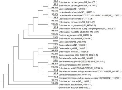
2012, Bahamdain et al. 2015). The 16S
rRNA analysis (Boye et al. 1999) further
supports this conclusion, as depicted in Fig. 8, illustrating the
phylogenetic tree of the PAH-degrading bacterial isolate (Isolate N°
1) and its relationship to other bacterial species based on the 16S
rRNA sequence.
Notably, (Isolate N° 1) appears closely related to
the strain Enterobacter asburiae. This
polyphasic appraisal combining phenotypic, chemotaxonomic and
genotypic analyses provides consistent evidence that Isolate N° 1
belongs to the Enterobacter asburiae species,
which includes strains proven to possess PAH biodegradation
capabilities.
Nucleotide sequence alignment between a Query
sequence and a Subject sequence (Enterobacter
asburiae strain JM-458 (NR_145647.1) 16S ribosomal RNA, partial
sequence):
Biodegradation Assay of Polycyclic Aromatic
Hydrocarbons
An experiment was conducted to assess the
biodegradation of three polycyclic aromatic hydrocarbons (PAHs) –
Naphthalene, Fluorene, and Anthracene – by Enterobacter asburiae strain N° 1. The results
from the biodegradation assay indicated residual concentrations of
1.05 ppb for Naphthalene, 0.516 ppb for Fluorene, and 0.862 ppb for
Anthracene after inoculation with 1 ml of bacterial suspension
(12*107 cfu/ml) of Enterobacter asburiae strain N°1.
Fig. 7.- Images depicting the complex growth of PAHs-degrading
bacteria on minimal salt medium plates supplemented with PAHs (1
µg/L). Specifically, (A) represents the complex growth culture
derived from the Kitchener Drain sample, (B) depicts the complex
growth culture from the Total gas station sample, and (C)
illustrates the complex growth culture originating from the Neamaa
restaurant sample
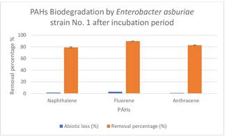
The experiment extended over 14 days at 35 °C with a
pH of 7.0. Considering the initial concentration of PAHs as 5 ppb,
the removal percentages for the three PAH compounds were determined
as 79 %, 89.7 %, and 82.8 %. The experiment was repeated twice, and
averages along with standard deviations were calculated. Table VII
presents the duplicate experiments, showcasing residual
concentrations of PAHs after a 14-day incubation period, including
calculated means and standard deviations.
In the control experiment, where no bacterial
presence was introduced, the residual concentrations after the
incubation period were measured as 4.92 ppb for Naphthalene, 4.85
ppb for Fluorene, and 4.95 ppb for Anthracene. Fig. 9 illustrates
the removal percentages for the three PAH compounds, taking into
account abiotic loss. The abiotic loss percentages for Naphthalene,
Fluorene, and Anthracene were recorded as 1.6 %, 3.1 %, and 1 %,
respectively.
Similar findings were reported by Belal et al. (2018) in a study using Paenibacillus sp. strain for testing the
biodegradation of Organochlorine Pesticides in aqueous solutions.
The study demonstrated efficient biodegradation ranging between 24.4
% and 98 % for different Organochlorine Pesticides after a two-week
incubation period. Additionally, Sarma et al.
(2019) developed a plant-microbes assisted remediation
technology involving bacterial strains, including Enterobacter asburiae, for polyaromatic
hydrocarbons (phenanthrene, anthracene, pyrene, and benzo[a]pyrene)
degradation and heavy metals (Cr, Ni, and Pb) removal in a
microcosmic experiment by bacterial strains used in two formulated
consortia includes ─ Cpm1 (Enterobacter cloacae
HS32, Brevibacillus reuszeri HS37, and
Stenotrophomonas sp. HS16) and Cpm2 (Acinetobacter junii HS29, Enterobacter aerogenes HS39 and Enterobacter asburiae HS22); Both the consortia
that were newly developed showed similar trends of metals removal
and PAHs degradation.
Enterobacter asburiae’s
biodegradation capability was further emphasized by El Gendy et al. (2020), who highlighted its potential for
the biodegradation and removal of Organochlorine pesticides residues
in an aqueous system, affirming the reduction of toxicity in the
tested organochlorine pesticides.
Numerous studies have explored various
microorganisms for their ability to degrade PAHs. Ahmed et al. (2010) found that Kocuria flava and Kocuria rosea
can grow on naphthalene, phenanthrene, and fluoranthene.
Haritash & Kaushik (2016) identified two bacterial species, Micrococcus luteus and Kocuria
rosea, capable of degrading low molecular weight PAHs, along
with three fungal species of Aspergillus
showing potential for PAH remediation. Goswami et al. (2018) demonstrated that Rhodococcus opacus bacteria could degrade a
mixture of PAHs, including Naphthalene, Phenanthrene, and
Fluoranthene.
The biodegradation mechanism of Enterobacter asburiae strain 1 in PAHs
degradation involves the utilization of PAHs as the sole carbon
source. This perspective has been discussed in various studies, such
as Igwo-Ezikpe et al. (2010), who isolated
bacterial strains from engine-oil polluted sites in Lagos, Nigeria.
These isolates, belonging to genera Micrococcus, Staphylococcus, Kurthia
sp., Acinetobacter, Pseudomonas, and Corynebacterium, exhibited varying rates of
growth on PAHs (anthracene, fluoranthene, and pyrene) as sole
sources of carbon and energy. The findings suggest that PAHs
degradation may be plasmid and/or chromosomally mediated, depending
on the bacterial isolate and the specific PAHs being degraded.
Igwo-Ezikpe et al. (2010) also revealed that
different compounds induce varied genetic changes in bacterial
isolates in response to stimuli.
Due to the possibility of formation of toxic
intermediate products during the biodegradation of Polycyclic
Aromatic Hydrocarbons, the evaluation of biodegradation ratio was
not sufficient to decide the effectiveness of the biodegradation
process; thus, the possible toxic impact of intermediate products
should be taken into account, and the toxicity of the degradation
intermediate products of the tested PAHs should be confirmed by the
experiment. Polycyclic Aromatic Hydrocarbons degrading bacterium
Enterobacter asburiae (Isolate N° 1) had the ability to biologically
degrade approximately 79 %, 89.7 % and 82.8 % of Naphthalene,
Fluorene and Anthracene active ingredients respectively with initial
concentration 5 ppb under optimum condition.
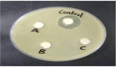
Toxicity of the remaining PAHs compounds in the
aqueous solution after 14 days of incubation with Enterobacter asburiae (Isolate N°1) was evaluated
using Bacillus subtilis as a target organism. The results of the
toxicity assessment showed that the PAHs compounds after 14 days of
incubation had no toxicity; consequently, there was no antibacterial
activity could be detected against B. subtilis
as a test organism. The obtained result was compared with
control treatment (active ingredients of PAHs without bacterial
activity) which revealed 100 % inhibition against B. subtilis growth under the same conditions as
showed in Fig. 10. This implied that the aqueous solutions spiked
with three PAHs compounds separately were detoxified after 14 days
of treatment with Enterobacter asburiae (Isolate N°1). Table VIII
represented the percentages % of the inhibition caused by
supernatant of PAHs compounds treated with bacterial isolates
against the target organism Bacillus subtilis
in comparison to the inhibition in the control experiment. Fig.
10 represented the control experiment and showed the 100 %
inhibition of Bacillus subtilis growth; and revealed the
bioremediation of PAHs without any antibacterial activity against
the target organism (Bacillus subtilis).
Polycyclic Aromatic Hydrocarbons have serious toxic and even lethal
effects on other microorganisms’ growth and metabolism.
The extensive toxicity assessment throughout the
degradation process would yield valuable insights into the
application of microbe-focused approaches for the bioremediation of
polycyclic aromatic hydrocarbons (PAHs) and ensures the safety of
organisms and the surrounding environment from potential hazardous
secondary metabolites resulting from the degradation process.
Shuttleworth & Cerniglia (1995) indicated that degradation
products of PAHs are, however, not necessarily less toxic than the
parent compounds; and therefore, toxicity assays need to be
incorporated into the procedures used to monitor the effectiveness
of PAH bioremediation.
Naphthalene treated by E. asburiae
(Isolate N°. 1)
14 Days
0
Fluorene treated by E. asburiae
(Isolate N°. 1)
14 Days
0
Anthracene treated by E. asburiae
(Isolate N°. 1)
14 Days
0
Control treatment (untreated
PAHs)
14 Days
100
Wulandari et al. (2021)
showed that the newly isolated Trametes polyzona
PBURU 12 demonstrated a high tolerance and potential for the
degradation of phenanthrene. The fungal isolate was able to tolerate
100 ppm of phenanthrene with 45 % relative growth and proved that
the biodegradation metabolites showed the absence of toxic compounds
as microbial viability tests using E. coli
and B. subtilis revealed that the
treated phenanthrene was less toxic than untreated phenanthrene;
also phytotoxicity and genotoxicity tests, using Vigna radiata and Allium
cepa, indicated that the treated phenanthrene was less toxic to
the plants and no mutagenic activity was found in the Ames test. In another study, Shanker et
al. (2017) examined the degradation of selected toxic PAHs (3-5
rings) using potassium zinc hexacyanoferrate (KZnHCF) nanocubes and
proved higher proficiency of the catalyst regarding PAHs degradation
into small and non-toxic by-products such as malealdehyde,
4-oxobut-2- enoic acid and o-xylene. On the other
hand, Wang et al. (2021) investigated
the accumulated pattern of the metabolites of phenanthrene
biodegradation by Rhodococcus qingshengii
strain FF and evaluated and their toxicity to Vibrio fischeri, effect on microbiota diversity
of farmland soil and influence on seed of wheat, and indicated that
the accumulated metabolites in later phase were more toxic to Vibrio fischeri, microbe and wheat seed response
to the different stages of phenanthrene metabolites indicated
pollution significantly decreased microbial richness and evenness of
farmland soil and lower germinal length, root length or root number
of wheat seed.
CONCLUSION
Three wastewater sources in Kafr El-Sheikh
governorate, Egypt, underwent testing to determine the presence of
Polycyclic Aromatic Hydrocarbons (PAHs) and were found to contain
varying concentrations of PAHs (ranging from 0.42 to 253.1 ppb). The
biodegradation of PAHs was distinctly observed using different
bacterial isolates in aqueous media. Numerous bacterial isolates with
the capability of degrading PAHs were obtained through an enrichment
technique from wastewater samples. These isolates were systematically
screened to select the most efficient ones, followed by
characterization, identification based on morphological and
biochemical characteristics, and 16S rRNA analysis. Subsequently,
these isolates were subjected to testing for their ability to
biodegrade PAHs. Enterobacter asburiae strain 1
demonstrated the capacity to biologically degrade approximately 79 %,
89.7 %, and 82.8 % of the active ingredients of Naphthalene, Fluorene,
and Anthracene, respectively. Furthermore, the results of the toxicity
assessment indicated that the PAHs compounds, after a 14- day
incubation period, underwent biodegradation, and secondary metabolites
were found to have no toxicity. Consequently, no antibacterial
activity could be detected against Bacillus
subtilis, employed as a test organism. This study advocates for
the use of Enterobacter asburiae strain for the
safe breakdown of PAHs found in industrial wastewater before reaching
surface water. The biological degradation of PAHs compounds offers a
more functional, cost-effective, and thermodynamically affordable
process for the elimination of such hazardous residues.
REFERENCES
Abdel-Shafy H I, Mansour M S 2016. A review on
polycyclic aromatic hydrocarbons: source, environmental impact, effect
on human health and remediation. Egypt J Petroleum
25(1): 107-123.Ahmed R Z, Ahmed N, Gadd G M 2010. Isolation of
two Kocuria species capable of growing on various polycyclic aromatic
hydrocarbons. Afric J Biotechnol 9(24):
3611-3617.Bahamdain L, Fahmy F, Lari S, Aly M
2015.Characterization of some Bacillus strains obtained from marine
habitats using different taxonomical methods. Life
Sci J 12: 58.Belal E S B, Shalaby M E, El-Gremi S M, Gad W A
2018. Biodegradation of organochlorine pesticides by Paenibacillus sp.
strain. Environ Engineering Sci 35(11):
1194-1205.Boffetta P, Jourenkova N, Gustavsson P 1997.
Cancer risk from occupational and environmental exposure to polycyclic
aromatic hydrocarbons. Cancer Causes Control
8(3): 444-472.Boye K, Høgdall E, Borre M 1999. Identification
of bacteria using two degenerate 16S rDNA sequencing primers. Microbiol Res 154(1): 23-26.Brunner W, Staub D, Leisinger T 1980. Bacterial
degradation of dichloromethane. Appl Environ Microb
40(5): 950-958.Cappuccino JG, Sherman N 1996. In Microbiology: ALaboratory Manual 4th edition,
Menlopark, CA, The Benjamin/Cunnings Publishing Company, Inc.Cerniglia C E 2003. Recent advances in the
biodegradation of polycyclic aromatic hydrocarbons by Mycobacterium
species. The utilization of bioremediation to reduce soil
contamination: problems and solutions: 51-73.Chang W, Um Y, Holoman T R P 2002. Polycyclic
aromatic hydrocarbon (PAH) degradation coupled to methanogenesis. Biotechnol Lett 24: 425-430.Chun J, Rhee M S, Han J I, Bae K S 2001.
Arthrobacter siderocapsulatus Dubinina & Zhdanov 1975. AL is a
later subjective synonym of Pseudomonas putida
(Trevisan 1889) Migula 1895AL. Intern J System
Evol Microb 51(1): 169-170.Dandie C E, Thomas S M, Bentham R H, McClure N
C 2004. Physiological characterization of Mycobacterium sp. strain 1B
isolated from a bacterial culture able to degrade
high‐molecular‐weight polycyclic aromatic hydrocarbons. J Appl Microbiol 97(2): 246-255.Dean-Ross D, Moody J D, Freeman J P, Doerge D
R, Cerniglia C E 2002. Metabolism of anthracene by Rhodococcus
species. FEMS Microbiol Lett 204:
205-211.Derbalah A S H, Belal E B, Massoud A H 2008.
Biodegradability of famoxadone by various microbial isolates in
aquatic systems. Land Contam Reclam 16(1): 13 -
23.Derbalah A S, Belal E B 2008. Biodegradation
kinetics of cymoxanil in aquatic system. Chem Ecol
24(3): 169-180.El Gendy S S, Belal E B, Sidkey N M, Abdelrazek
M A, Metwaly M M, Gad WA 2020. Monitoring and bioremediation of
organochlorine pesticides in surface water with Enterobacter asburiae.
Spanish J Agricult Res 18(4): 12.Fredslund L, Sniegowski K, Wick L Y, Jacobsen C
S, De Mot R, Springael D 2008. Surface motility of polycyclic aromatic
hydrocarbon (PAH)-degrading mycobacteria. Res
Microbiol 159(4): 255-262.Gallego J L R, Loredo J, Llamas J F, Vázquez F,
Sánchez J 2001. Bioremediation of diesel-contaminated soils. Intern Biodeter Biodeg 47: 249-255.Gerhardt P, Murry E, Costilow R, Nester E, Wood
W, Kreig N, Philips GB 1981. In Manual of Methods of General
Bacteriology. Washington, DC, American Society for Microbiol.Gordon R E, Haynes W C, Pang C H N 1973. The genus Bacillus (N°. 427). Agricultural Research
Service, US Department of Agriculture, Washington DC, USA.Goswami L, Manikandan N A, Dolman B,
Pakshirajan K, Pugazhenthi G 2018. Biological treatment of wastewater
containing a mixture of polycyclic aromatic hydrocarbons using the
oleaginous bacterium Rhodococcus opacus. J Cleaner
Prod 196: 1282-1291.Guo Y, Huang E, Yuan C, Zhang L, Yousef A E
2012. Isolation of a Paenibacillus sp. strain and structural
elucidation of its broad-spectrum lipopeptide antibiotic. Appl Environ Microbiol 78: 3156.Gupte A, Tripathi A, Patel H, Rudakiya D, Gupte
S 2016. Bioremediation of polycyclic aromatic hydrocarbon (PAHs): a
perspective. Open Biotechnol J 10: 363-378.
doi: 10.2174/1874070701610010363.Haritash A K, Kaushik C P 2009. Biodegradation
aspects of polycyclic aromatic hydrocarbons (PAHs): a review. J Hazardous Mat 169(1-3): 1-15.Haritash A K, Kaushik C P 2016. Degradation of
low molecular weight polycyclic aromatic hydrocarbons by
microorganisms isolated from contaminated soil. Intern J Environ Sci 6(5): 808-819.Hauka F I, Belal E, Selim M A A, Gad A I 2014.
Bioremediation of chemical pollutants- contaminated water
a-biodecolorization of crystal violet contaminated water by Pseudomonas geniculata. J Agric
Chem Biotech 5(5): 165-176.Hodgeson J W 1990. Determination of Polycyclic
Aromatic Hydrocarbons in Drinking Water by Liquid-liquid Extraction
and HPLC with Coupled Ultraviolet and Fluorescence Detection: Test
Method 550. US Environmental Protection Agency.Igwo-Ezikpe M N, Okpuzor J, Awodele O,
Nwaokorie F O, Fowora M A, Akinbo M O 2010. Prevalence of polycyclic
aromatic hydrocarbons (PAHs) degrading bacteria in contaminated
tropical soil in Lagos, Nigeria: involvement of plasmid in
degradation. Inter J Biol Chem Sci 4(6):
2133-2145.Juhasz A L, Stanley G A, Britz M L 1997.
Microbial degradation and detoxification of high molecular weight
polycyclic aromatic hydrocarbons by Stenotrophmonas maltophilia strain
VUN 10,003. Lett Appl Microbiol 25:
396-401.Krieg N R, Holt J G 1984. Bergey’s manual of
systematic bacteriology. Yi Hsien Publishing Co.Lee B K, Vu V T 2010. “Sources, distribution
and toxicity of polyaromatic hydrocarbons (PAHs) in particulate
matter,” in Air Pollution, (London:IntechOpen): 99-122.Logan N A 2005. Bacillus
anthracis, Bacillus cereus and other
aerobic endospore-forming bacteria. In Boriello
SP, Murray PR, Funke G Eds, Topley and Wilson’s Microbiology and
Microbial Infections. Bacteriology 2:
922-952.Massoud A H, Derbalah A S, Belal E S B 2008.
Microbial detoxification of metalaxyl in aquatic system. J Environ Sci 20(3): 262-267.McDevitt S 2009. Methyl red and voges-proskauer
test protocols. Amer Soc Microb 8.Mojiri A, Zhou J L, Ohashi A, Ozaki N,
Kindaichi T 2019. Comprehensive review of polycyclic aromatic
hydrocarbons in water sources, their effects and treatments. Sci Total Environ 2019: 133971. doi:
10.1016/j.scitotenv.2019.133971.Muller R H, Thibault M A, Deomenico P 1996.
Enumeration and isolation of soil and sediment bacteria capable of
mineralizing aromatic hydrocarbons. Can J Microbiol
42: 759-767.Nessim G D, Seita M, Plata D L, O’Brien K P,
Hart A J, Meshot E R, Thompson C V 2011. Precursor gas chemistry
determines the crystallinity of carbon nanotubes synthesized at low
temperature. Carbon 49(3): 804-810.Okere U, Semple K 2012. Biodegradation of PAHs
in ‘pristine’soils from different climatic regions. J Bioremed Biodegrad S1: 006. doi:
10.4172/2155-6199.S1-006.Opuene K, Agbozu I E, Ekeh L E 2007.
Identification of perylene in sediments: occurrence and diagenetic
evolution. Intern J Environ Sci Techn 4(4):
457-462.Peng R H, Xiong A S, Xue Y, Fu X Y, Gao F, Zhao
W, Yao Q H 2008. Microbial biodegradation of polyaromatic
hydrocarbons. FEMS microbiol reviews 32(6):
927-955.Rangaswami G, Bagyaraj D J 1993. Microbial
biotechnology. In Agricultural Microbiol. New Delhi, India,Prentice
Hall of India Pvt. Ltd: 389-405.Rengarajan T, Rajendran P, Nandakumar N,
Lokeshkumar B, Rajendran P, Nishigaki I 2015. Exposure to polycyclic
aromatic hydrocarbons with special focus on cancer. Asian Pac J Trop Biomed 5: 182-189.Rubin H 2001. Synergistic mechanisms in
carcinogenesis by polycyclic aromatic hydrocarbons and by tobacco
smoke: a bio-historical perspective with updates. Carcinogenesis 22(12): 1903-1930.Samanta S K, Singh O V, Jain R K 2002.
Polycyclic aromatic hydrocarbons: environmental pollution and
bioremediation. Trends Biotechnol 20:
243-248.Sarma H, Sonowal S, Prasad M N V 2019.
Plant-microbiome assisted and biochar-amended remediation of heavy
metals and polyaromatic compounds- a microcosmic study. Ecotox Environ Safety: 176: 288-299.Seeley H W, Vandemark P J 1981. Microbes in
Action-A Laboratory Manual of Microbiol. San Francisco, Freeman and
Company: 388.Shanker U, Jassal V, Rani M 2017. Degradation
of toxic PAHs in water and soil using potassium zinc hexacyanoferrate
nanocubes. J Environ Manag 204:
337-348.Sharma A, Krishna V, Kaur P, Rayal R 2015.
Characterization and Screening of Various MulberryVarieties Throgh Morpho-Biochemical
Characteristics. J Glob Biosci 4(1):
1186-92.Shuttleworth K L, Cerniglia E 1995.
Environmental aspects of PAH biodegradation. Appl
Biochem Biotechnol 54: 291-302.Singleton I, Mihailova A, Rush D 2009. Methods
for the isolation and identification of alkane- utilising bacteria
from petroleum reservoir fluid and subsurface samples. In Timmis K N ed. Microbiology of Hydrocarbons,
Oils, Lipids and Derived Compounds. Springer-Verlag Berlin Heidelberg:
29-54.Suman S, Sinha A, Tarafdar A 2016. Polycyclic
aromatic hydrocarbons (PAHs) concentration levels, pattern, source
identification and soil toxicity assessment in urban traffic soil of
Dhanbad. India. Sci Total Environ 545: 353-360.
doi: 10.1016/j.scitotenv.2015.12.061.Tejeda-Agredano M C, Gallego S, Niqui-Arroyo J
L, Vila J, Grifoll M, Ortega-Calvo J J 2011.Isolation of PAH-degrading bacteria from a
microbial consortium. Biodegradation 22:
705-711.Tolosa I, Bayona J M, Albaigés J 1996.
Aliphatic and polycyclic aromatic hydrocarbons and sulfur/oxygen
derivatives in northwestern Mediterranean sediments: spatial and
temporal variability, fluxes, and budgets. Environ
Sci Techn 30(8): 2495-2503.Uppstad H, Osnes G H, Cole K J, Phillips D H,
Haugen A, Mollerup S 2011. Sex differences in susceptibility to PAHs
is an intrinsic property of human lung adenocarcinoma cells. Lung Cancer 71(3): 264-270.US EPA (US Environmental Protection Agency).
(2003). National Primary Drinking Water Standards.Wang Y, Nie M, Diwu Z, Chang F, Nie H, Zhang
B,Yin Q 2021. Toxicity evaluation of the metabolites derived from the
degradation of phenanthrene by one of a soil ubiquitous PAHs-degrading
strain Rhodococcus qingshengii FF. J Hazardous Mat
415: 125657.Westley P A, Schindler D E, Quinn T P,
Ruggerone G T, Hilborn R 2010. Natural habitat change, commercial
fishing, climate, and dispersal interact to restructure an Alaskan
fish metacommunity. Oecologia 163(2):
471-484.World Health Organization 2006. Guidelines for
drinking-water quality, WHO, Geneva, Switzerland, 3rd edition.Wu B, Zhang R, Cheng S P, Ford T, Li A M, Zhang
X X 2011. Risk assessment of polycyclic aromatic hydrocarbons in
aquatic ecosystems. Ecotoxicology 20(5):
1124-1130.Wulandari R, Lotrakul P, Punnapayak H, Amirta
R, Kim S W, Prasongsuk S 2021. Toxicity evaluation and biodegradation
of phenanthrene by laccase from Trametes polyzona PBURU 12. 3 Biotech 11: 1-11.Yu H 2002. Environmental carcinogenic
polycyclic aromatic hydrocarbons: photochemistry and phototoxicity.
J Environ Sci Health Part C 20(2):
149-183.Yu H, Huang G H, Xiao H, Wang Q 2005.
Development of ROC curves for prediction of PAHs biodegradability. J Environ Informatics 5 (1): 36-45.Zhang J, Wang ZT, Yu HM, Ma Y 2013.
Paenibacilluscatalpae sp. nov., isolated from the rhizosphere soil of
Catalpa speciosa. Int J Syst Evol Microbiol 63:
1776.Zheng H, Xing X, Hu T, Zhang Y, Zhang J, Zhu G
et al. 2018. Biomass burning contributed most
to the human cancer risk exposed to the soil-bound PAHs from Chengdu
Economic Region, western China. Ecotoxicol Environ
Saf 159: 63-70. doi: 10.1016/j.ecoenv.2018.04.065.Zou Y, Yin H, Tan Q, Chen Y, Lv G, Hou X 2011.
Polycyclic aromatic hydrocarbons (PAHs) pollution recorded in annual
rings of gingko (Gingko biloba L.): regression
analysis and comparison to other pollutants. Microchem J 98(2): 303-306.
Fig.1.- Chromatogram of three PAHs compounds - Anthracene,
Fluorene, and Naphthalene - in water sample collected from Kitchener
drain before incubation periodFig. 2. - Chromatogram of three PAHs compounds - Anthracene,
Fluorene, and Naphthalene - in water sample collected from Kitchener
drain after incubation period.Fig. 3. - Chromatogram of three PAHs compounds - Anthracene,
Fluorene, and Naphthalene - in water sample collected from Neamaa
restaurant before incubation periodFig. 4.- Chromatogram of three PAHs compounds - Anthracene,
Fluorene, and Naphthalene - in water sample collected from Neamaa
restaurant after incubation periodFig. 5.- Chromatogram of three PAHs compounds - Anthracene,
Fluorene, and Naphthalene - in water sample collected from total
gas station before incubation periodFig 6.-. Chromatogram of three PAHs compounds - Anthracene,
Fluorene, and Naphthalene - in water sample collected from total
gas station aftier incubation periodFig. 7.- Images depicting the complex growth of PAHs-degrading
bacteria on minimal salt medium plates supplemented with PAHs (1
µg/L). Specifically, (A) represents the complex growth culture
derived from the Kitchener Drain sample, (B) depicts the complex
growth culture from the Total gas station sample, and (C)
illustrates the complex growth culture originating from the Neamaa
restaurant sample
BIODEGRADATION OF POLYCYCLIC AROMATIC HYDROCARBONS
WITH ENTEROBACTER ASBURIAE
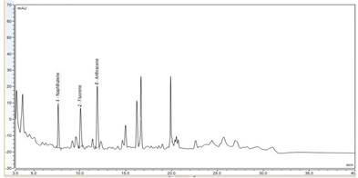 Fig.1.- Chromatogram of three PAHs compounds - Anthracene,
Fluorene, and Naphthalene - in water sample collected from Kitchener
drain before incubation period
Fig.1.- Chromatogram of three PAHs compounds - Anthracene,
Fluorene, and Naphthalene - in water sample collected from Kitchener
drain before incubation period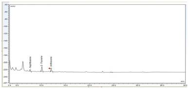 Fig. 2. - Chromatogram of three PAHs compounds - Anthracene,
Fluorene, and Naphthalene - in water sample collected from Kitchener
drain after incubation period.
Fig. 2. - Chromatogram of three PAHs compounds - Anthracene,
Fluorene, and Naphthalene - in water sample collected from Kitchener
drain after incubation period.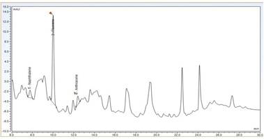 Fig. 3. - Chromatogram of three PAHs compounds - Anthracene,
Fluorene, and Naphthalene - in water sample collected from Neamaa
restaurant before incubation period
Fig. 3. - Chromatogram of three PAHs compounds - Anthracene,
Fluorene, and Naphthalene - in water sample collected from Neamaa
restaurant before incubation period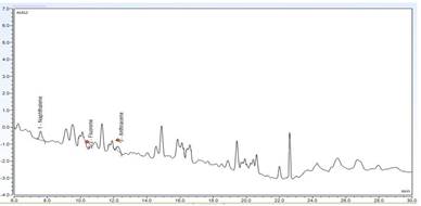 Fig. 4.- Chromatogram of three PAHs compounds - Anthracene,
Fluorene, and Naphthalene - in water sample collected from Neamaa
restaurant after incubation period
Fig. 4.- Chromatogram of three PAHs compounds - Anthracene,
Fluorene, and Naphthalene - in water sample collected from Neamaa
restaurant after incubation period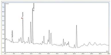 Fig. 5.- Chromatogram of three PAHs compounds - Anthracene,
Fluorene, and Naphthalene - in water sample collected from total
gas station before incubation period
Fig. 5.- Chromatogram of three PAHs compounds - Anthracene,
Fluorene, and Naphthalene - in water sample collected from total
gas station before incubation period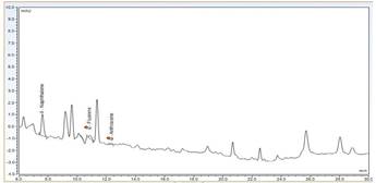 Fig 6.-. Chromatogram of three PAHs compounds - Anthracene,
Fluorene, and Naphthalene - in water sample collected from total
gas station aftier incubation period
Fig 6.-. Chromatogram of three PAHs compounds - Anthracene,
Fluorene, and Naphthalene - in water sample collected from total
gas station aftier incubation period
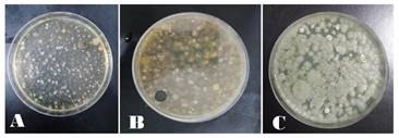 Fig. 7.- Images depicting the complex growth of PAHs-degrading
bacteria on minimal salt medium plates supplemented with PAHs (1
µg/L). Specifically, (A) represents the complex growth culture
derived from the Kitchener Drain sample, (B) depicts the complex
growth culture from the Total gas station sample, and (C)
illustrates the complex growth culture originating from the Neamaa
restaurant sample
Fig. 7.- Images depicting the complex growth of PAHs-degrading
bacteria on minimal salt medium plates supplemented with PAHs (1
µg/L). Specifically, (A) represents the complex growth culture
derived from the Kitchener Drain sample, (B) depicts the complex
growth culture from the Total gas station sample, and (C)
illustrates the complex growth culture originating from the Neamaa
restaurant sample